The following abstract is drawn from a recently published paper in Journal of Pathology Informatics | ScienceDirect.com by Elsevier. We invite you to read the full paper and join the conversation, become a member of the Pathology News community to share your thoughts, ask questions, and engage with others around this work.
Authors: Falah Jabara, Pasquale Lombardib, Aria Torkpourb, Masoud Tafavvoghic, Per Niklas Benzler Waalerc, Sigve Andersend, Erna-Elise Paulsene, Mette Pøhlf, Lill-Tove Rasmussen Busunda, Tom Donnemd, Elin Richardsena, David J. Pinatob, Mehrdad Rakaeeg
Abstract
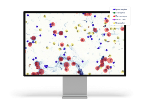
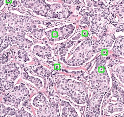
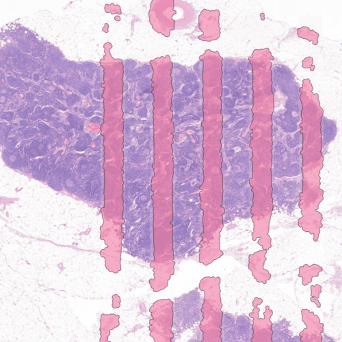
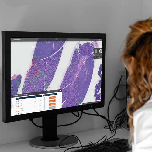
Accurate nuclei detection and classification in hematoxylin and eosin (H&E) whole-slide images (WSIs) is a key task in computational pathology, particularly for quantitative analysis of the tumor microenvironment. However, this task remains highly challenging due to variations in nuclei morphology, staining procedures, scanners, organs, magnifications, and WSI artifacts. In addition, many existing pipelines rely on computationally demanding architectures and post-processing procedures, making gigapixel WSI analysis time-consuming. In this work, CellPrior-Net (CP-Net) is proposed, an efficient nuclei detection and classification pipeline that utilizes a lightweight convolutional neural network architecture and hematoxylin (H) channel as prior information to enhance nuclei-aware feature learning. Extensive benchmarking was conducted against state-of-the-art pipelines on eight public and private datasets (total:~10.4 M nuclei) obtained from different organs, scanners, magnifications, and clinical centers. Experimental results demonstrate that CP-Net achieves comparable performance while significantly reducing inference time. Furthermore, CellQuant-Net was introduced—an end-to-end nuclei quantification pipeline—that integrates a quality assessment model to exclude regions with artifacts, followed by CP-Net cell detection and classification. The pipeline is publicly available on GitHub, and provides a potentially efficient and scalable framework for downstream computational pathology applications.
Read the full article: CellPrior-net: Prior-guided nuclei detection and classification for H&E whole-slide images – ScienceDirect
- aDep. of Clinical Pathology, University Hospital of North Norway, Tromsø, Norway
- bDep. of Surgery and Cancer, Imperial College London, London, United Kingdom
- cDepartment of Medical Biology, UiT The Arctic University of Norway, Tromsø, Norway
- dDep. of Clinical Medicine, UiT The Arctic University of Norway, Tromsø, Norway
- eDep. of Pulmonology, University Hospital of North Norway, Tromsø, Norway
- fDep. of Clinical Medicine, University of Copenhagen, Copenhagen, Denmark
- gDep. of Cancer Genetics, Oslo University Hospital, Oslo, Norway